Body Protection Compound-157
BPC-157 | Pentadecapeptide | PL 14736 | PL-10 | Bepecin
Mechanism of Action
BPC-157 is a synthetic pentadecapeptide derived from a protective protein found in human gastric juice. Its mechanisms of action are multifaceted and have been studied extensively in over 100 animal studies. A central aspect of its activity involves upregulation of growth factor expression, including VEGF (vascular endothelial growth factor), EGF (epidermal growth factor), and their receptors. This pro-angiogenic activity helps explain its remarkable wound-healing and tissue-repair properties observed across multiple tissue types.
BPC-157 also interacts with the nitric oxide (NO) system in a complex, context-dependent manner. It can rescue NO production when it is pathologically inhibited and can attenuate excessive NO when it is overproduced, suggesting a modulatory rather than unidirectional effect. Research by Sikiric et al. has demonstrated that BPC-157 interacts with the dopaminergic system and may counteract both the acute and chronic effects of dopaminergic agents, pointing to direct CNS activity.
At the gastrointestinal level, BPC-157 maintains mucosal integrity by promoting granulation tissue formation and angiogenesis within lesion sites. It has shown cytoprotective effects against NSAID-induced gastric damage, ethanol-induced lesions, and stress ulcers in numerous rodent models. The peptide appears to modulate the FAK-paxillin pathway, which is critical for cell migration and adhesion during wound repair.
Key Research Findings
- Sikiric et al. (2011) reviewed decades of research showing BPC-157 heals esophageal, gastric, duodenal, and colonic lesions in rodent models, with efficacy comparable to or exceeding standard treatments.
- Chang et al. (2011) demonstrated BPC-157 accelerated healing of transected Achilles tendons in rats by promoting tendon fibroblast outgrowth and VEGF expression.
- Seiwerth et al. (2014) showed BPC-157 promoted angiogenesis in a chick embryo CAM assay and accelerated cutaneous wound healing in diabetic rodent models.
- Pevec et al. (2010) found BPC-157 improved healing of medial collateral ligament injuries in rats with increased biomechanical strength at the repair site.
- Sikiric et al. (2018) demonstrated BPC-157 interacts with the NO system, rescuing impaired healing in L-NAME-treated animals and counteracting excessive NO in L-arginine models.
References
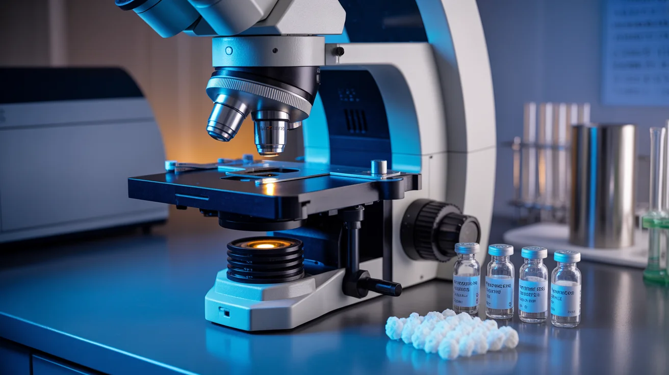
Dosage in Research
In rodent studies, BPC-157 is typically administered at 10 mcg/kg or 10 ng/kg, delivered intraperitoneally or locally at the injury site. Oral administration has also been studied for gastrointestinal applications. No human clinical trial data is currently published.
Storage & Handling
Store lyophilized powder at -20C, protected from light. Reconstituted solution should be refrigerated at 2-8C and used within 14-21 days. Use bacteriostatic water for reconstitution.
Frequently Asked Questions
What is BPC-157?
BPC-157 is a synthetic 15-amino-acid peptide derived from a naturally occurring protein in human gastric juice called Body Protection Compound. It has been studied extensively in animal models for its broad tissue-protective and healing properties.
What types of tissue repair has BPC-157 been studied for?
Animal studies have investigated BPC-157 in tendon, ligament, muscle, bone, skin, corneal, and gastrointestinal tissue repair. It has shown pro-healing effects across all these tissue types, which researchers attribute to its pro-angiogenic and growth factor modulatory activity.
Are there human clinical trials for BPC-157?
As of current literature, BPC-157 has been studied primarily in animal models and in vitro systems. While its safety profile in animal studies has been favorable (no reported toxicity at therapeutic doses), published human clinical trial data remains limited.
How does BPC-157 relate to TB-500?
BPC-157 and TB-500 (thymosin beta-4) are often studied in parallel due to their complementary tissue-repair mechanisms. BPC-157 works primarily through angiogenesis and growth factor modulation, while TB-500 promotes cell migration via actin polymerization regulation. This is the rationale behind blend products like the Wolverine Blend.
Source Body Protection Compound-157 from Research Vials
98%+ purity • US-accredited independent lab COA • Domestic cold-chain shipping